2026
Vesicle Segmentation
A public reconstruction of my Harris Lab vesicle segmentation work, using synthetic microscopy volumes instead of lab data.
- Role
- Sole engineer
- Stack
- Python, Zarr, 3-D U-Net, GitHub Actions
What it is
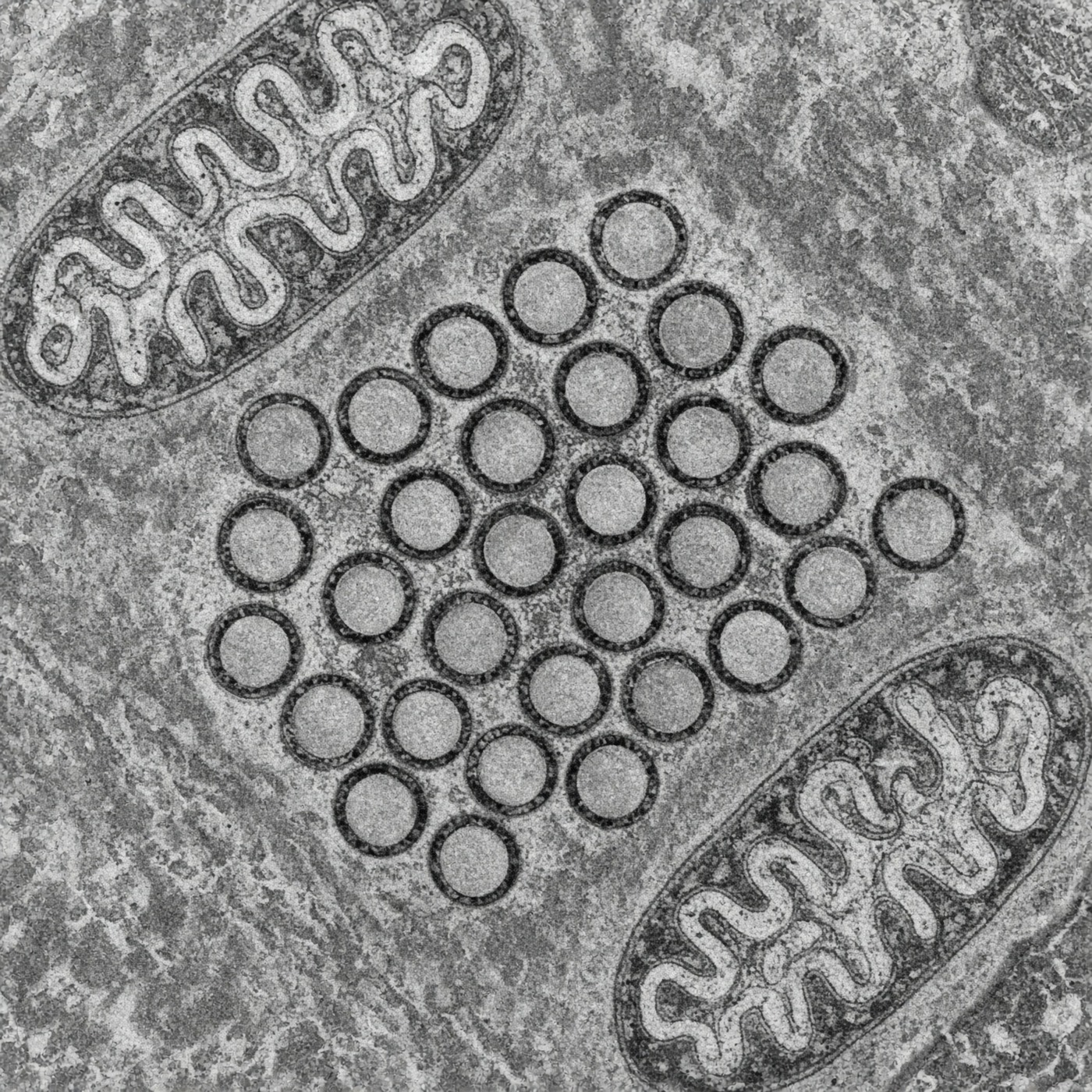
At the Harris Lab at UT Austin, I worked on detecting vesicles in large electron microscopy volumes. The lab data cannot be published, so this repository reconstructs the workflow using simulated volumes with 40 to 60 nm vesicles and mitochondria-like distractors. It contains no lab data or lab code.
How it runs
- Volumes are processed in chunks instead of being loaded entirely into memory.
- Adjacent chunks overlap during inference so objects crossing a chunk boundary can be reconciled.
- Training and test data are split by volume and checked for overlap.
- The 3D model is compared with a simple intensity threshold as a baseline.
Results
On the synthetic volumes, the model detects lower-intensity vesicles that the threshold baseline misses. Chunked inference also allows the pipeline to process volumes larger than available memory. These results come from a short training run on simulated data and are not Harris Lab results.
Limits
- All public results use synthetic volumes rather than lab electron microscopy data.
- Training is intentionally short and uses small volumes. The repository demonstrates the pipeline rather than a fully tuned segmentation model.